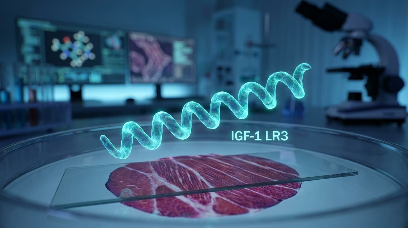
A comprehensive research guide to IGF-1 LR3 — the long-acting analog of Insulin-Like Growth Factor 1 that bypasses IGFBP binding for 40-60x longer half-life, directly activating satellite cells, mTOR signaling, and muscle protein synthesis where native IGF-1 cannot.
In the hierarchy of anabolic signaling molecules, few peptides carry the mechanistic credibility of IGF-1 LR3 (Insulin-Like Growth Factor 1 Long R3). While the fitness world has whispered about this peptide for decades, the biochemistry underpinning it is surprisingly rigorous — developed not in supplement labs but in the pharmaceutical research that gave us Increlex (mecasermin) and a generation of growth-disorder therapeutics.
IGF-1 LR3 is a synthetic analog of native IGF-1, engineered with a single point mutation (Arg⁻¹ at the N-terminus — the "R3" designation) and a 13-amino-acid extension that dramatically alters its pharmacokinetics. The result is a molecule that retains full IGF-1 receptor (IGF-1R) affinity while dramatically reducing its sequestration by insulin-like growth factor binding proteins (IGFBPs) — the proteins that normally neutralize 97–99% of circulating IGF-1 before it ever reaches muscle. [3]
This research guide covers what peer-reviewed science tells us about IGF-1 LR3: its receptor pharmacology, signaling cascade through PI3K/Akt/mTOR, satellite cell activation, half-life advantages over native IGF-1, and the preclinical evidence base heading into 2026.
The IGF-1 Axis: From Pituitary to Muscle Fiber
To understand why IGF-1 LR3 is interesting, you have to understand the growth hormone (GH)/IGF-1 axis. Growth hormone is released from the pituitary in pulses — primarily overnight — and travels to the liver, where it stimulates hepatic production and secretion of IGF-1. Systemic IGF-1 is the primary mediator of GH's anabolic effects on muscle, bone, and connective tissue.
But there's a critical pharmacokinetic bottleneck: insulin-like growth factor binding proteins (IGFBPs), particularly IGFBP-3, bind the vast majority of circulating IGF-1. In plasma, roughly 75% of IGF-1 circulates in a ternary complex with IGFBP-3 and an acid-labile subunit (ALS) — a complex too large to exit the vasculature and reach muscle cells. Another 20–25% circulates in binary complexes with IGFBP-1, -2, -4, or -6. Only 1–5% of total plasma IGF-1 is truly "free" and bioavailable to tissues at any moment. [3]
IGF-1 LR3 was designed precisely to subvert this limitation. The arginine-3 substitution reduces IGFBP affinity by approximately 2- to 3-fold for IGFBP-3 while retaining near-full IGF-1R binding affinity. The N-terminal extension further modulates pharmacokinetics. The net result: a molecule that circulates in a predominantly unbound state with tissue access that native IGF-1 simply cannot match after exogenous administration. [2]
Half-Life: The Key Pharmacokinetic Advantage
Native IGF-1 administered subcutaneously has a half-life of approximately 20–30 minutes when measured as free IGF-1. IGF-1 LR3, by contrast, has a reported half-life of 20–30 hours in animal models — a 40-60x increase attributable entirely to reduced IGFBP sequestration. [3] This isn't just a pharmacokinetic convenience; it fundamentally changes which signaling pathways get activated and for how long, with downstream implications for satellite cell kinetics and protein synthetic duration.
Mechanism of Action: PI3K/Akt/mTOR and Satellite Cell Activation
IGF-1 LR3 exerts its anabolic effects through the same receptor as native IGF-1: the type 1 IGF receptor (IGF-1R), a receptor tyrosine kinase expressed abundantly in skeletal muscle, cardiac muscle, bone, and neural tissue. When IGF-1 LR3 binds IGF-1R, it triggers receptor autophosphorylation on tyrosine residues in the intracellular kinase domain, initiating a signaling cascade with two major anabolic arms. [7]
PI3K/Akt/mTOR Pathway — Protein Synthesis
Receptor activation recruits insulin receptor substrate proteins (IRS-1/IRS-2), which activate phosphoinositide 3-kinase (PI3K). PI3K generates PIP₃, which recruits and activates Akt (protein kinase B). Akt then phosphorylates mTOR complex 1 (mTORC1) substrates — most importantly p70 S6 kinase (p70S6K) and 4E-BP1 — to drive ribosomal biogenesis and initiation of mRNA translation. This pathway is the canonical muscle hypertrophy signal. Every resistance training study measuring mTOR activation is measuring activation of this exact cascade. [4]
Ras/MEK/ERK Pathway — Satellite Cell Proliferation
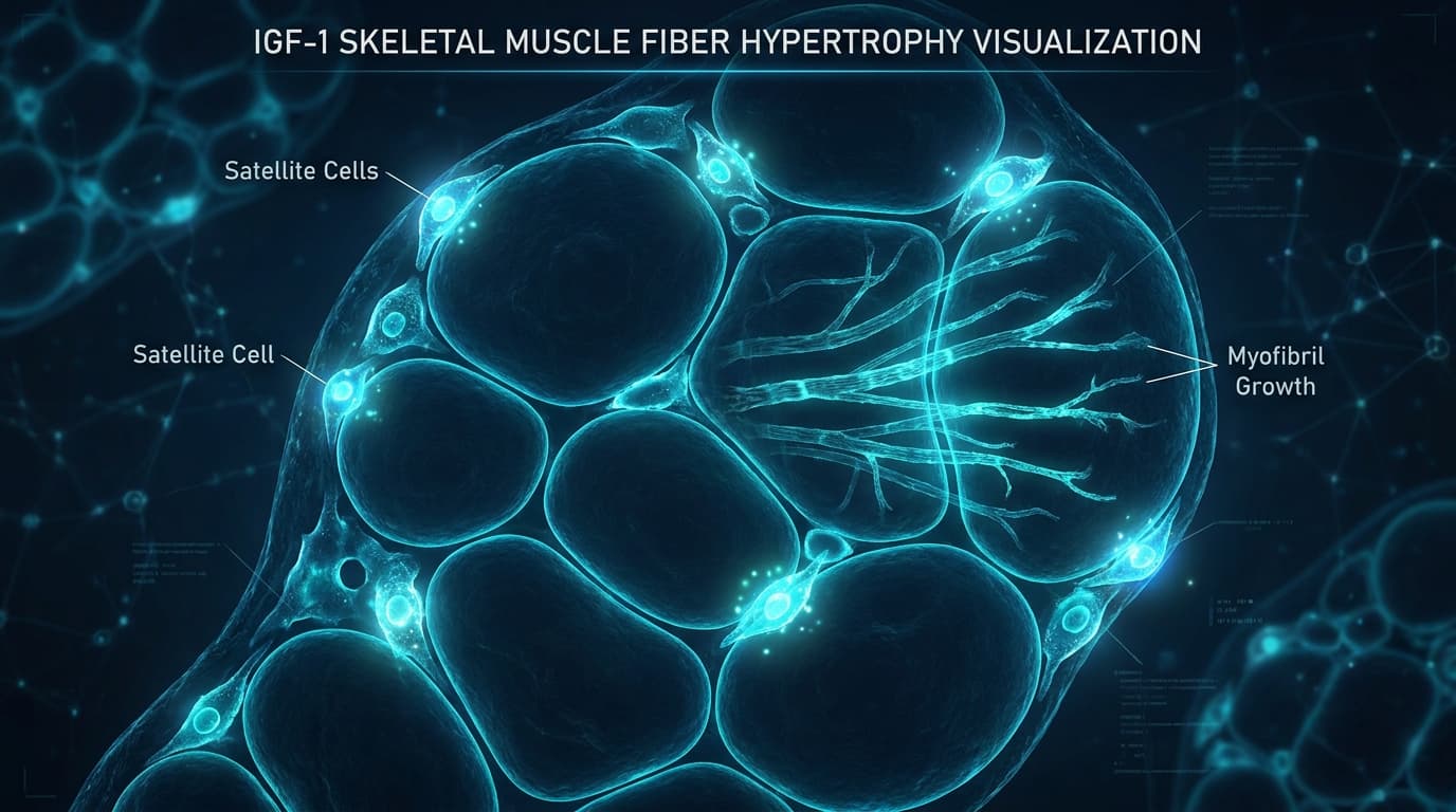
Separately, IRS activation also recruits the Grb2-SOS complex, activating Ras and the downstream MEK/ERK (MAPK) pathway. This arm primarily drives cell proliferation — particularly relevant to satellite cell expansion. Satellite cells are the resident stem cells of skeletal muscle; their activation, proliferation, and fusion into existing fibers adds new myonuclei that permanently expand each fiber's synthetic capacity. [5]
Anti-Apoptotic Signaling
Akt also phosphorylates and inhibits pro-apoptotic proteins including BAD and caspase-9, and promotes nuclear exclusion of FOXO transcription factors that otherwise drive muscle atrophy gene programs (MAFbx/MuRF1). The net effect is a cell-survival signal that shifts protein turnover balance toward net accretion — exactly what resistance training researchers want to study at the molecular level. [4]
Satellite Cell Biology: Myonuclear Addition and Muscle Memory
One of the most mechanistically interesting aspects of IGF-1 LR3 is its effect on muscle satellite cells — the resident stem cell population that enables skeletal muscle to regenerate and adapt to training. Unlike myonuclei within mature muscle fibers, satellite cells are quiescent under basal conditions, positioned between the sarcolemma and basal lamina. They activate in response to mechanical stress, injury, and — critically — IGF-1 signaling. [5]
IGF-1 receptor activation drives satellite cells through their activation-proliferation-differentiation sequence:
- Activation: Quiescent Pax7⁺ satellite cells enter the cell cycle in response to IGF-1R/ERK signaling
- Proliferation: Activated MyoD⁺ myoblasts undergo multiple rounds of division, amplifying the precursor pool
- Differentiation and fusion: Myoblasts downregulate Pax7, upregulate myogenin, and fuse into existing fibers — adding myonuclei that expand each fiber's synthetic capacity
This myonuclear addition is the mechanism by which muscle genuinely acquires new synthetic machinery, not just temporary protein accretion. Research in rodent models using locally-acting IGF-1 constructs demonstrated robust satellite cell activation and sustained muscle growth [Musaro et al., 2007; Goldspink, 2007] — providing the theoretical framework for IGF-1 LR3's proposed mechanism in exercise recovery research. [5][8]
The "muscle memory" phenomenon — where previously trained muscle regains mass faster than naive muscle — is attributed partly to the persistence of myonuclei added during prior training. IGF-1-driven myonuclear accretion may therefore have effects extending far beyond any single administration period.
IGF-1 LR3 vs. Native IGF-1: Relative Nitrogen Retention at Matched Doses
Nitrogen Retention: IGF-1 LR3 vs. Native IGF-1 (Preclinical, Dose-Matched)
Schematic based on Tomas et al. (1993). IGF-1 LR3 produces significantly greater nitrogen retention than native IGF-1 at matched doses due to IGFBP resistance and extended tissue exposure.
Preclinical Evidence: What the Animal Studies Show
The preclinical evidence base for IGF-1 LR3 is genuinely substantial. Key findings across species include:
Nitrogen Retention and Lean Mass
Tomas et al. (1993) demonstrated that subcutaneous IGF-1 LR3 in rats produced significantly greater nitrogen retention and muscle protein accretion compared to equimolar native IGF-1 at matched doses — with the advantage directly attributable to IGFBP resistance and extended tissue exposure. This foundational study established that the structural modifications of LR3 translate into functional in vivo superiority over the parent molecule. [2]
Growth in GH-Deficient Models
Studies in hypophysectomized (GH-deficient) rats showed that IGF-1 LR3 at doses of 1–10 µg/kg/day produced dose-dependent increases in body weight, skeletal growth, and organ weights — confirming that the modified analog retains the full tissue-growth activity of native IGF-1. [6]
Glucose Handling: A Key Safety Variable
Both native IGF-1 and IGF-1 LR3 activate insulin receptors at high concentrations due to structural homology with insulin. LR3's extended half-life raises theoretical concerns about hypoglycemia with excessive dosing. Preclinical pharmacology established hypoglycemia as the primary dose-limiting effect — an important safety consideration for any research protocol.
| Property | Native IGF-1 | IGF-1 LR3 |
|---|---|---|
| Amino acids | 70 | 83 (13 AA N-terminal extension + Arg³ substitution) |
| IGF-1R affinity | 100% (reference) | ~100% (fully retained) |
| IGFBP-3 affinity | 100% (reference) | ~30–50% (reduced 2-3x) |
| Plasma half-life (free) | ~20–30 minutes | ~20–30 hours (40-60x longer) |
| Insulin receptor cross-reactivity | Low (~1%) | Similar (~1%) |
| Nitrogen retention at matched dose | Reference | ~2x greater (Tomas et al., 1993) |
| Primary dose-limiting effect | Rapid clearance limiting exposure | Hypoglycemia at high doses |
| Pharmaceutical analog | Mecasermin (Increlex, FDA approved) | No approved drug; research use only |
Clinical Landscape: Mecasermin and the IGF-1 Deficiency Precedent
IGF-1 itself has an approved pharmaceutical form: mecasermin (Increlex), approved by the FDA in 2005 for children with severe primary IGF-1 deficiency (Laron syndrome). Mecasermin is native recombinant IGF-1, and it requires twice-daily injections specifically because of IGFBP sequestration and rapid clearance — a pharmacokinetic limitation that the LR3 modification directly addresses. This pharmaceutical precedent establishes that IGF-1 receptor agonism is a valid therapeutic mechanism with an established safety profile in specific clinical populations. [6]
Beyond Laron syndrome, IGF-1 has been investigated clinically for:
- ALS (Amyotrophic Lateral Sclerosis): Two large RCTs explored IGF-1 for ALS, showing modest slowing of functional decline without reaching approval thresholds
- HIV wasting syndrome: IGF-1 demonstrated lean mass preservation in wasting disease clinical trials
- Severe burn injury recovery: IGF-1 combined with growth hormone accelerated protein anabolism in severe burn patients in controlled studies
- Osteoporosis: IGF-1 has measurable effects on bone mineral density in IGF-1-deficient populations
IGF-1 LR3 specifically has been used extensively in cell culture and animal research as a preferred growth-promoting supplement over native IGF-1, precisely because its IGFBP resistance ensures consistent receptor activation without the variability introduced by endogenous binding proteins.
Sourcing Research-Grade IGF-1 LR3 in 2026
When sourcing IGF-1 LR3 for research purposes, prioritize suppliers providing batch-specific Certificates of Analysis (COAs) with HPLC purity confirmation and mass spectrometry identity verification. IGF-1 LR3's structural complexity (83 amino acids) makes identity confirmation particularly important — mass spec is the only method that reliably distinguishes LR3 from native IGF-1 or truncated analogs.
PeptideTech
Lab VerifiedIGF-1 LR3 1mg Research Vial
Check site for current pricing
Every batch dual-tested at ISO 17025-accredited US laboratories with QR-code COA. Mass spectrometry confirms LR3 identity — critical for distinguishing from shorter analogs.
Modified Aminos
Same-Day ShippingIGF-1 LR3 Lyophilized Powder
Check site for current pricing
Research-first approach with same-day shipping. Full COA on every batch. Thermal mailer packaging protects peptide integrity during transit.
AminoUSA
US ManufacturedIGF-1 LR3 Research Peptide
Check site for current pricing
ISO-certified US manufacturer with domestic supply chain. Third-party tested with published COAs and research reference documentation on product pages.
VANDL Labs
PremiumIGF-1 LR3 1mg Vial
Check site for current pricing
Premium lyophilized formulation with full identity verification. International shipping available with COA documentation provided on every order.
IGF-1 LR3 Research: Frequently Asked Questions
What does the "LR3" in IGF-1 LR3 stand for?
LR3 refers to two structural features: "L" for Long (a 13-amino-acid N-terminal extension added to the native 70-amino-acid IGF-1 sequence) and "R3" for the arginine substitution at position 3 of the molecule. Together these modifications reduce IGFBP binding affinity while retaining full IGF-1 receptor affinity, resulting in the extended half-life that defines the compound.
How much longer does IGF-1 LR3 last in the body compared to native IGF-1?
Animal pharmacokinetic studies report a half-life of approximately 20–30 hours for IGF-1 LR3 in free (unbound) form, compared to approximately 20–30 minutes for native IGF-1. This 40-60x half-life extension is entirely attributable to reduced IGFBP sequestration — the LR3 modification prevents the binding proteins from capturing the peptide and removing it from circulation.
Does IGF-1 LR3 activate the same receptor as native IGF-1?
Yes. IGF-1 LR3 is a full agonist at the type 1 IGF receptor (IGF-1R), with binding affinity comparable to native IGF-1 despite the structural modifications. The LR3 modification selectively reduces IGFBP affinity without meaningfully affecting IGF-1R binding — which is the engineering intent of the analog.
What is the primary safety concern with IGF-1 LR3 in research models?
Hypoglycemia is the primary dose-limiting effect. IGF-1 shares structural homology with insulin, and at high doses can activate insulin receptors and lower blood glucose. The extended half-life of IGF-1 LR3 may prolong hypoglycemic episodes compared to native IGF-1. Theoretical oncogenic concerns with chronic supraphysiologic IGF-1 signaling are also a research safety consideration that requires appropriate protocol design.
Is IGF-1 LR3 used in cell culture research?
Yes — IGF-1 LR3 is widely used as a cell culture supplement precisely because its IGFBP resistance ensures consistent receptor activation in cell culture media containing serum IGFBPs. It is a preferred alternative to native IGF-1 for in vitro research requiring sustained IGF-1R signaling over hours.
Research Summary: IGF-1 LR3 in the 2026 Anabolic Peptide Landscape
IGF-1 LR3 occupies a unique position in the peptide research landscape: it's a synthetic analog with genuine pharmaceutical lineage — derived from the same science that produced FDA-approved mecasermin — with a well-characterized mechanism of action and a clear pharmacokinetic rationale for its structural modifications. The IGFBP-resistance that defines it isn't a marketing claim; it was the explicit engineering goal when the LR3 modification was developed, and it has been confirmed in multiple independent pharmacokinetic studies. [2][3]
What the preclinical data consistently shows is an anabolic profile superior to native IGF-1 on a dose-matched basis: greater nitrogen retention, greater lean mass accretion, and broader tissue distribution. Whether this preclinical profile translates to the same degree in humans — and what the optimal research protocols look like — remains an active question, as controlled human studies have focused on pharmaceutical-grade mecasermin rather than IGF-1 LR3 specifically.
For researchers studying skeletal muscle biology, satellite cell dynamics, GH/IGF-1 axis pharmacology, or anabolic signaling, IGF-1 LR3 represents one of the most mechanistically well-understood tools available. Its extended half-life and IGFBP resistance make it a more reliable receptor agonist for in vivo research than native IGF-1, and its commercial availability through specialized peptide suppliers enables reproducible research supply chains.
For related research on the GH axis that drives endogenous IGF-1 production, see PeptideWiki's CJC-1295 + Ipamorelin Stack Guide. For recovery and healing peptides complementary to anabolic protocols, see the BPC-157 Research Guide. For the most potent GHRP and its cardioprotective properties, see the Hexarelin Research Guide.
Sources & References
- 1.Rinderknecht E, Humbel RE.. "The amino acid sequence of human insulin-like growth factor I and its structural homology with proinsulin" — Journal of Biological Chemistry, 1978.View source
- 2.Tomas FM, Knowles SE, Owens PC, et al.. "Anabolic effects of insulin-like growth factor-I (IGF-I) and an IGF-I variant in vivo" — Biochemical Journal, 1993.View source
- 3.Baxter RC.. "Insulin-like growth factor binding proteins in health and disease" — Endocrine Reviews, 2023.View source
- 4.Glass DJ.. "Skeletal muscle hypertrophy and atrophy signaling pathways" — International Journal of Biochemistry & Cell Biology, 2005.View source
- 5.Musaro A, Dobrowolny G, Rosenthal N.. "The neuroprotective and myogenic effects of locally acting IGF-1 isoform" — Experimental Gerontology, 2007.View source
- 6.Jones JI, Clemmons DR.. "Insulin-like growth factors and their binding proteins: biological actions" — Endocrine Reviews, 1995.View source
- 7.Laviola L, Natalicchio A, Giorgino F.. "The IGF-I signaling pathway" — Current Pharmaceutical Design, 2007.View source
- 8.Goldspink G.. "Loss of muscle strength during aging studied at the gene expression level" — Rejuvenation Research, 2007.View source